Stitching of 3x3 FoVs
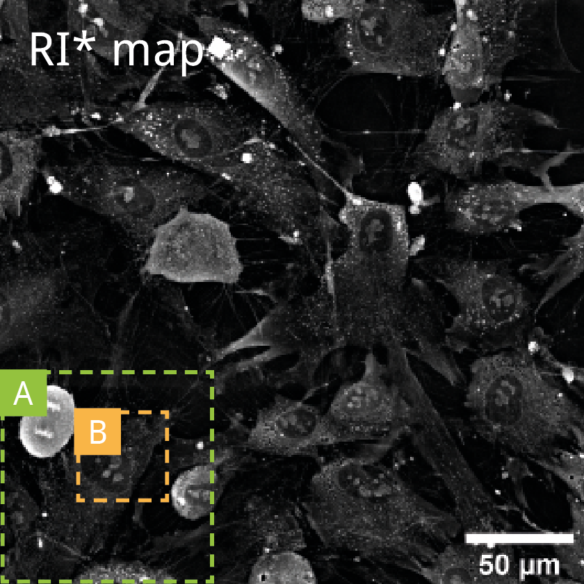
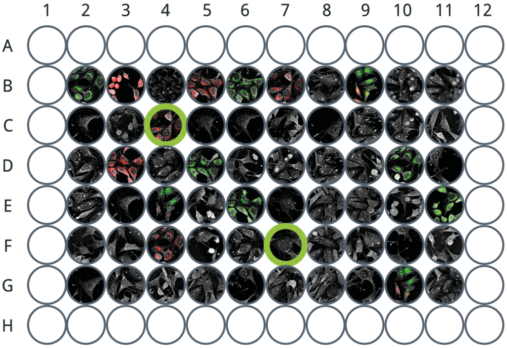
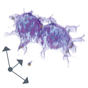
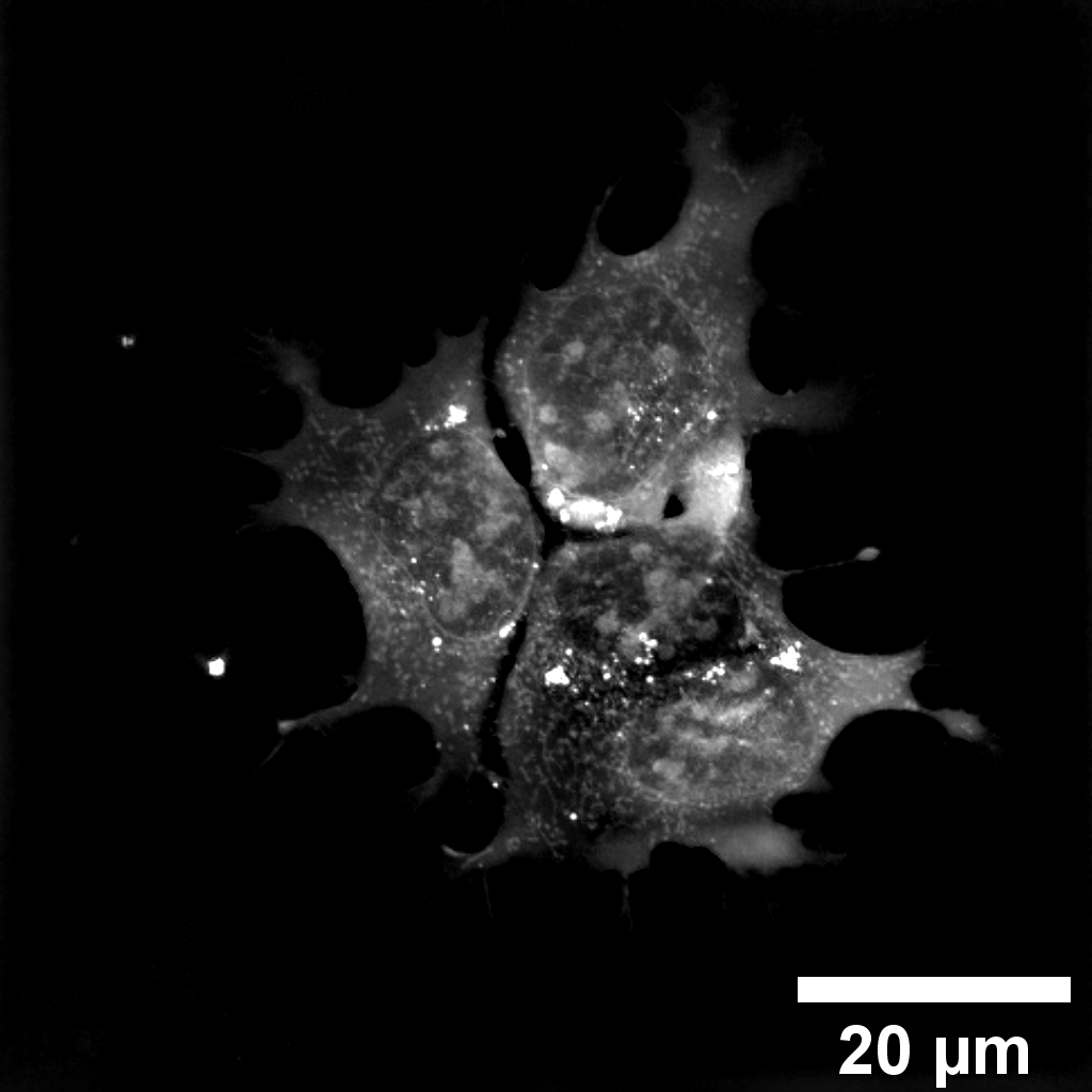
Cell population
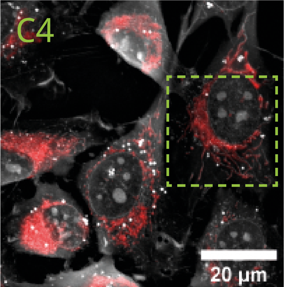
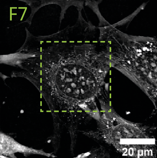
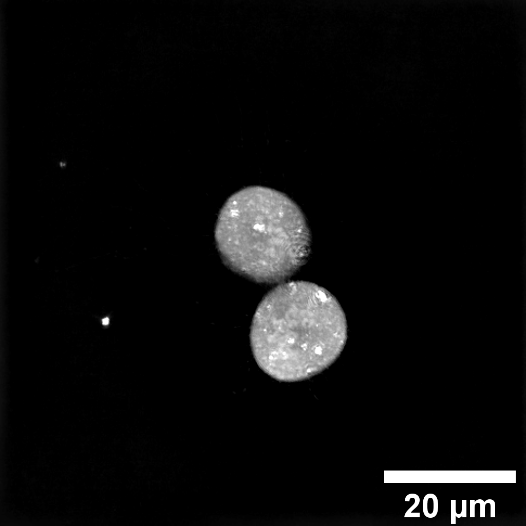
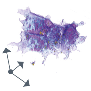
Single FoV
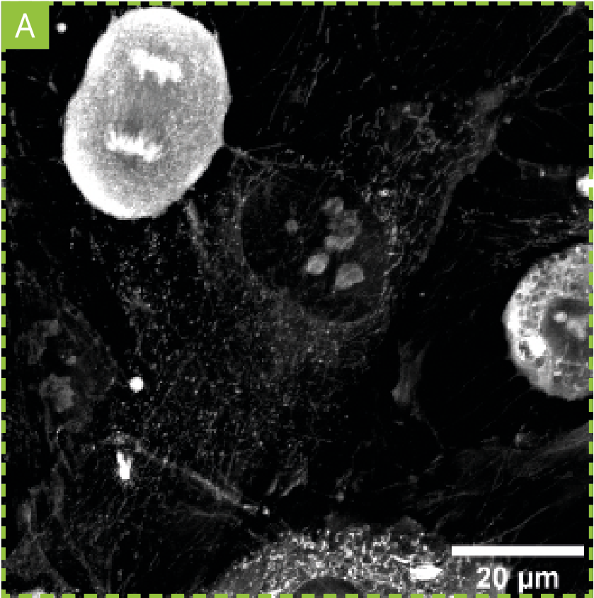
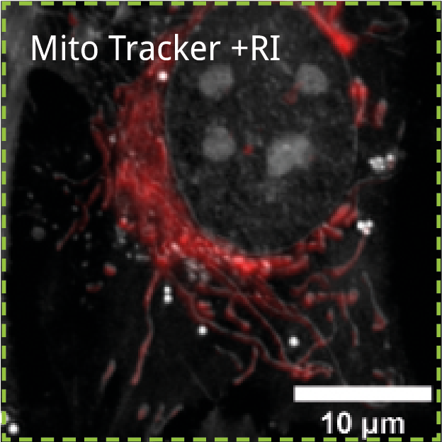
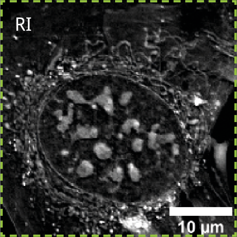
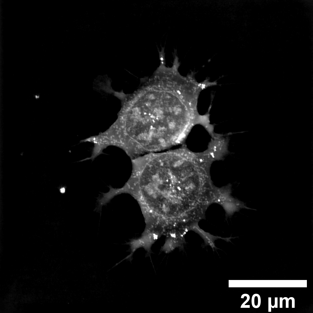
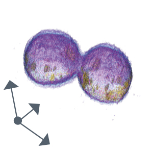
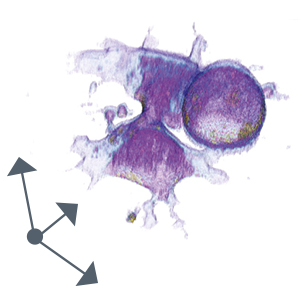
Single cell
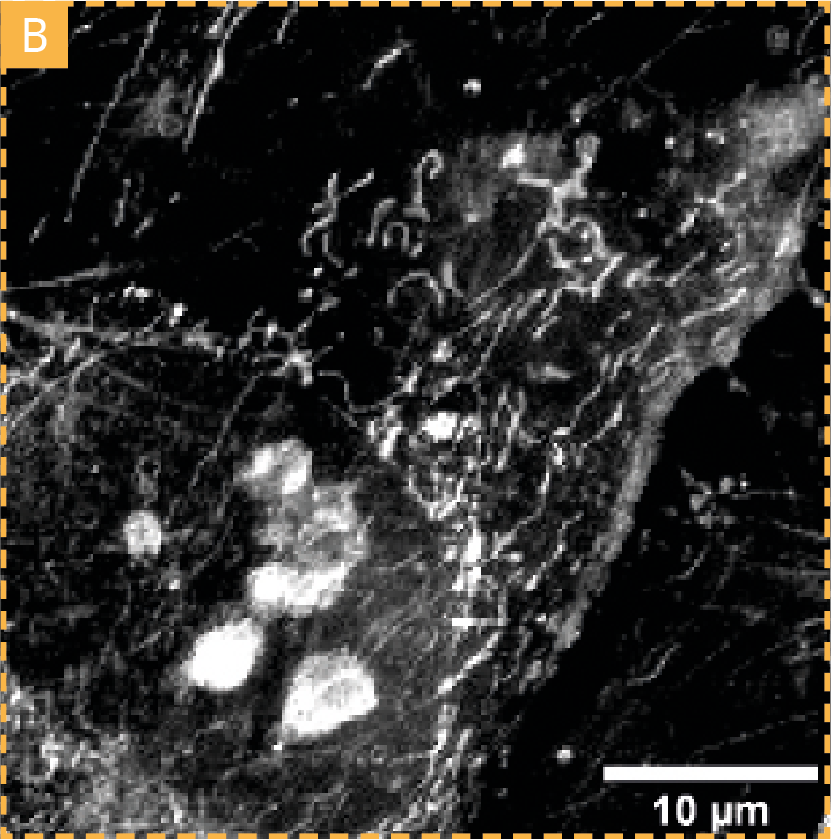
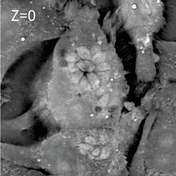
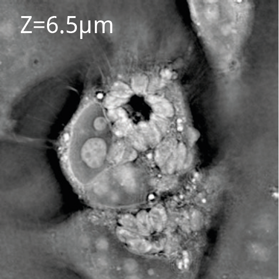
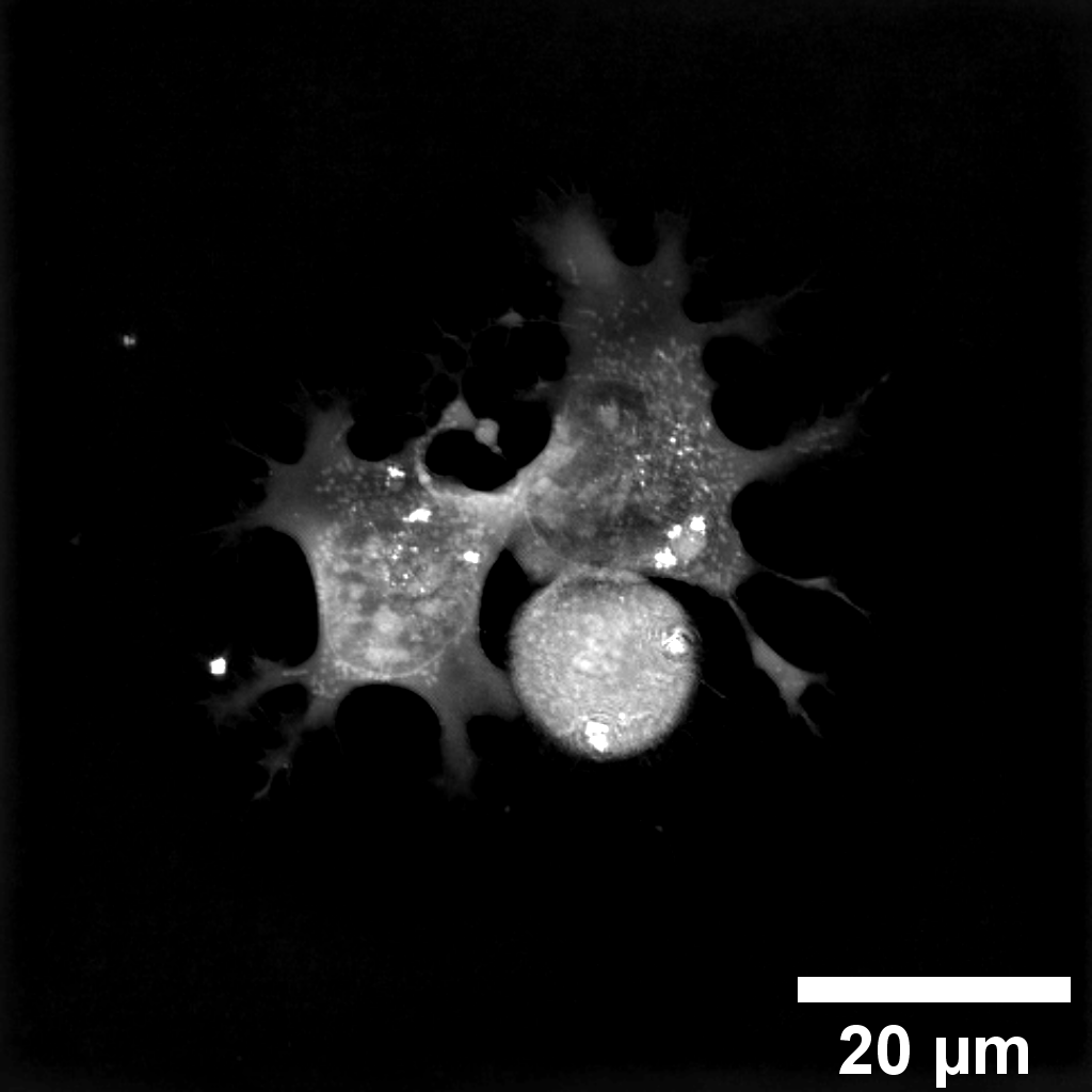
Zoom into FoV
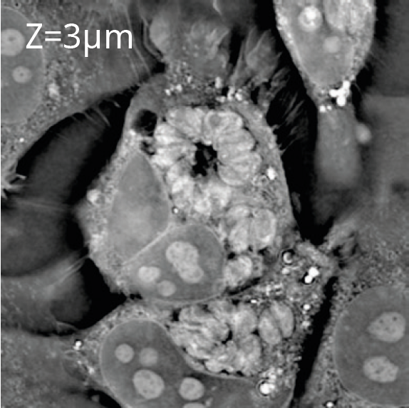
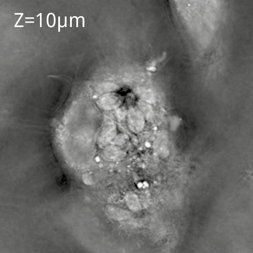
Organelle ecosystem
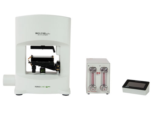
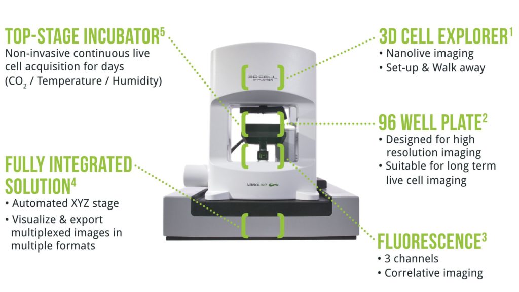
Long-term live cell incubation
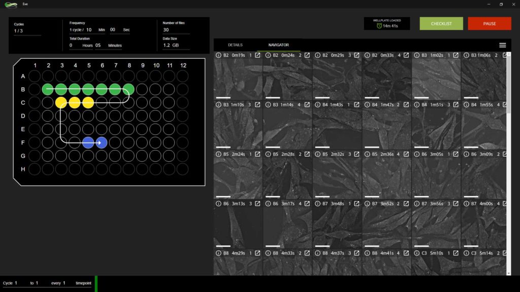
Walk-away with automation
Nanolive’s CX-A automates data acquisition, thus delivering new insights and understanding into biological processes.
Nanolive’s completely redesigned intuitive user-interface enables first-time users to set-up experiments in just a few minutes and walk away, while the CX-A automatically collects the images. In addition to improving data significance, multiple imaging regimens can be programmed within the same plate allowing users to run different applications in parallel. A real-time preview allows the user to navigate through the data at any time while offering an overview of the experimental protocol. The 3D dataset output can then be exported with just a few clicks to multiple data formats for analysis.
See what you have been missing
Technical Specifications
| 1,3 Illumination source | Holotomography (HT): Class 1 laser low power (λ=520 nm, sample exposure 0.2 mW/mm2) Epifluorescence: High speed switchable <100 μs, Lifetime >20’000 hours |
|---|---|
| 1,3 Resolution | HT: 3D: x,y: 200 nm; z: 400 nm Epifluorescence: x,y: ~ 400 nm (depending on channel) |
| 1,3 Field-of-view (FoV) | HT: Single FoV* / 2×2 FoV / 3×3 FoV *Single FoV: RI: 90x90x30 μm; Epifluorescence: 90×90 μm |
| 1,3 Channels | HT: Up to 8 organelles simultaneously Epifluorescence: DAPI + FitC + TritC | FitC + TritC + Cy5 | DAPI + FitC + TritC/Cy5 |
| 1,3 Imaging modalities | Automated: 3D HT | 3D HT + Epifluorescence | 4D HT time-lapse | 4D HT + Epifluorescence time-lapse |
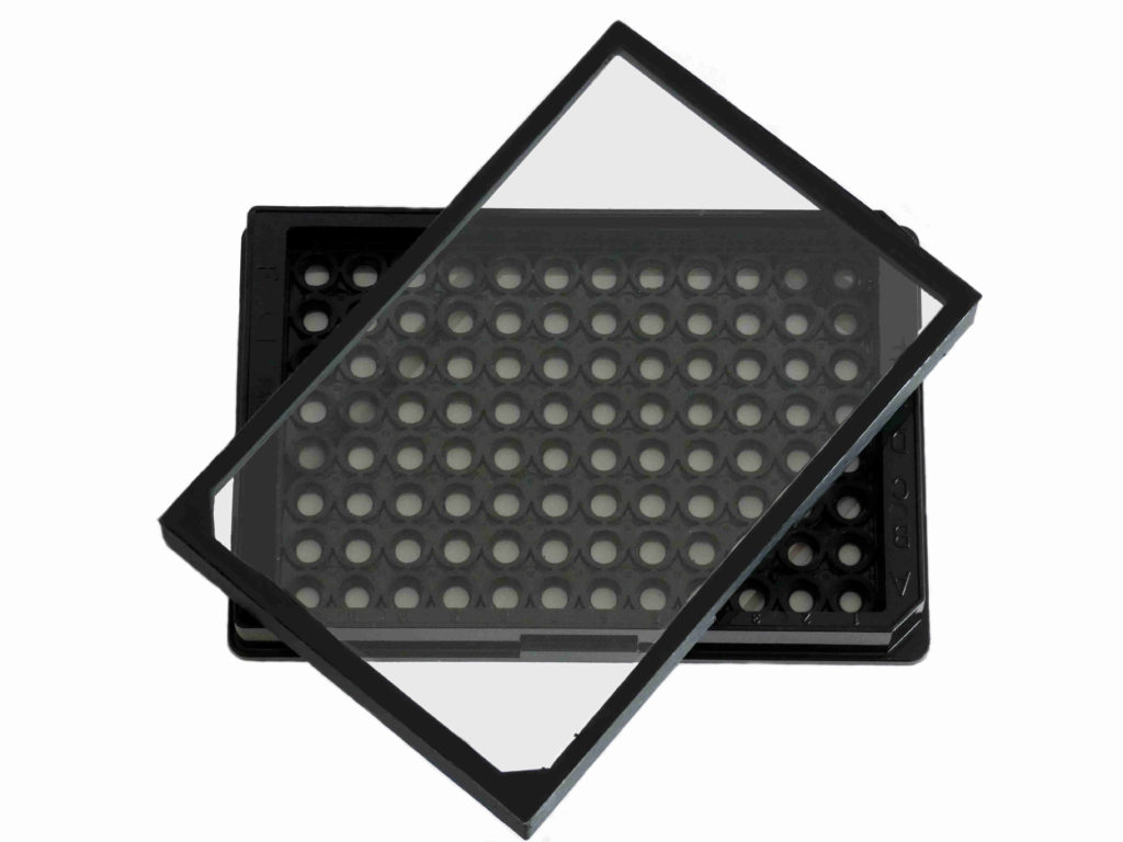
| 2 Sample holder | Nanolive’s 96 well format, designed for high precision imaging with optical quality glass bottom and lid |
| 1,3 Autofocus | High precision label-free autofocus for stable long-term observations in all imaging modalities |
| 5 Incubator stage-top | Tokaihit stage top incubator: CO2 concentration range: 5% – 20% (±0.1%); Humidity: ~ 95%; Sample temperature: 30-40°C (±0.3°C) |
| 1,3 Camera | USB 3.0 CMOS Sony IMX174 sensor / Quantum Efficiency (typical) 70 % (at 545 nm) / Dark Noise (typical) 6,6 e¯ / Dynamic Range (typical) 73,7 dB |
| 1,3 Microscope Objective | Dry objective / 60x magnification / NA 0.8 |
| 1-5 Weight | ~30Kg |