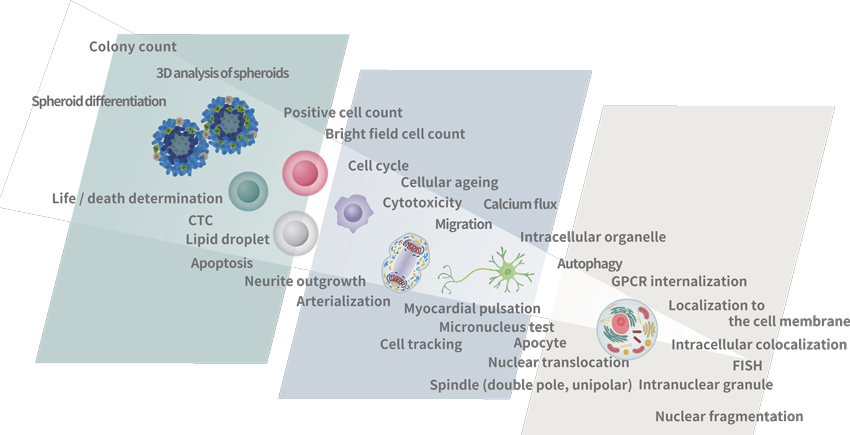
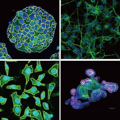
Enables measurement of spheroids, colonies and tissue sections
- No need to remove cells from culture dish, in contrast to traditional flow cytometry
- Nipkow spinning disk confocal technology allows high-speed yet gentle 3D image acquisition
- Rich feature extraction to facilitate sophisticated cellular image analysis
- Wide field of view and tiling capability enable easy imaging of large specimen
Enables analysis of time lapse and live cell
- High precision stage incubator and low phototoxicity of our confocal makes the analysis of time lapce and live cell are possible
- Max.20fps option for fast time lapce*1
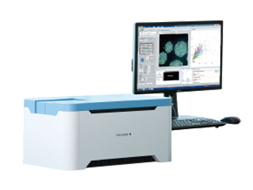
High quality image and the similar operability to traditional flow cytometer
- Feature data and statistical graphs displayed in real-time with image acquisition
- Usuable high quality image as confocal micorscope image.
- Easy to trace back to the original image from a graph spot, and make repetitive measurements
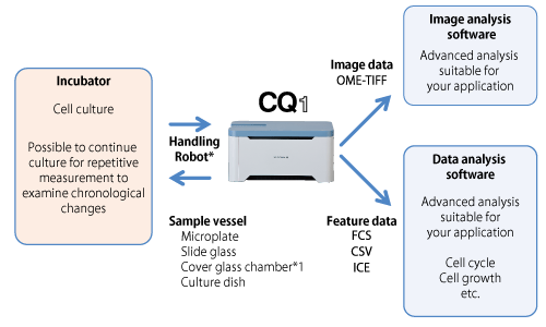
Open platform
- Connectable with external systems via handling robot*2
- Expandable to integrated system as image acquisition and quantification instrument
- FCS/CSV/ICE data format readable by third-party data analysis software
- A variety of cell culture and sample dishes are applicable
Compact footprint, light weight bench-top device; no need for darkroom
| CQ1 | General fluorescent imaging | Flow cytometry | |
|---|---|---|---|
| Cell removal/suspension treatment | Not necessary | Not necessary | Necessary |
| Cell image confirmation | Possible | Possible | Not possible |
| Display feature data and graphs in real-time with imaging | Possible | Depends on devices | Possible |
| 3D data measurement | Possible | Not possible | Not possible |
| Time lapse | Possible | Not possible | Not possible |
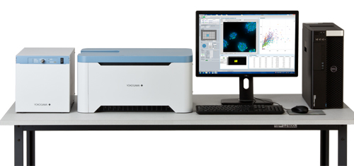
Multiple functions fully integrated in a compact box
Compact design contains fully integrated multiple functions to offer easy-to-handle confocal imaging system, without a need for complicated system integration; you only need to set a sample and run the software. User-friendly interface and versatile functions support your measurement and analysis.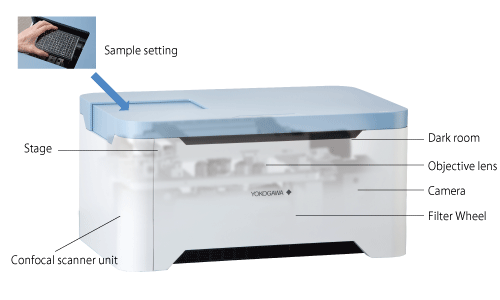
Principles of the Microlens-enhanced Nipkow Disk Scanning Technology
A Nipkow spinning disk containing about 20,000 pinholes and a subsidiary spinning disk containing the same number of microlenses to focus excitation laser light into each corresponding pinhole are mechanically fixed on a motor, and very rapidly rotated. As a result, high-speed raster scan of the excitation lights on the specimen can be achieved.The pinhole and microlenses are arranged on each disk in our proprietary design to optimize raster scan. Multi-beam scanning not only increases scanning speed, but also results in significantly lower photobleaching and phototoxicity, because multi-beam excitation needs only low level of laser power on the specimen to fully excite fluorescence.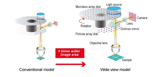
System integration with CQ1
Example of setup
| Item | Specifications |
|---|---|
| Optics | Microlens enhanced dual wide Nipkow disk confocal, Phase contrast (Optional add-on) |
| Laser/Filter | Laser : Choose 2-4 lasers from 405/488/561/640nm, 10-position Filter wheel (built-in) |
| Camera | sCMOS 2560×2160pixel, 16.6×14.0mm |
| Objective lens | Max.6 lenses (Dry: 2x, 4x, 10x, 20x, 40x Long working distance: 20x, 40x Phase contrast: 10x, 20x ) |
| Sample vessel | Microplate (6, 24, 96, 384 well), Slide glass, Cover glass chamber*1, Dish (35, 60mm*1) |
| XY stage | High-precision XY stage, designated resolution 0.1µm |
| Z focus | Electric Z motor, designated resolution 0.1µm |
| Autofocus | Laser autofocus, Software autofocus |
| Feature data | Number of cells/cellular granules, Intensity, Volume, Surface area, Area, Perimeter, Diameter, Sphericity, Circularity, etc |
| Data format | Image : 16bit TIFF file (OME-TIFF), PNG Numerical data : FCS, CSV, ICE |
| Workstation | Measurement and analysis workstation |
| Size/weight | Main unit : 600×400×298mm 38kg Utility box : 275×432×298mm 18kg |
| Environment | 15 – 30oC、20 – 70%RH No condensation |
| Power consumption | Main unit and Utility box : 100-240VAC 800VAmax, Workstation : 100-240VAC 650VAmax |