CellVoyager™ CQ1 Benchtop High-Content Analysis System
Home » Cell Imaging & Invivo Imaging » High Content Confocal Imaging » Benchtop CQ1 Confocal System

CellVoyager CQ1 enables 3D imaging and quantification of live cell clusters, such as spheroids within a 3D culture vessel, as they are, keeping the cells intact. CellVoyager CQ1 exports feature data in general formats which are readable by various third-party software for advanced data analysis. It is possible to construct a fully customized CellVoyager CQ1-based system by integrating with external systems*1, via robot for culture dish handling.
CellVoyager CQ1 System Highlights
| Excitation laser wavelength | 405 nm, 488 nm, 561 nm, 640 nm |
| Illumination source | Laser |
| Objective lens | 2x to 60x (Dry, Phase contrast, Long working distance) |
| Camera | High-sensitivity sCMOS camera |
| Autofocus | Laser autofocus, Software autofocus |
| Software | CellPathfinder |
Enables measurement of spheroids, colonies, and tissue sections

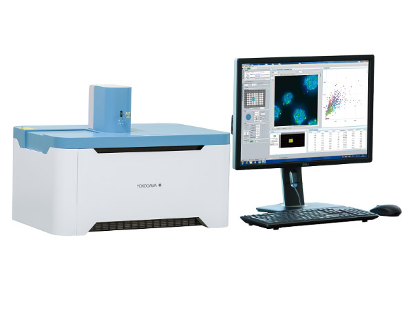
- No need to remove cells from the culture dish, in contrast to traditional flow cytometry
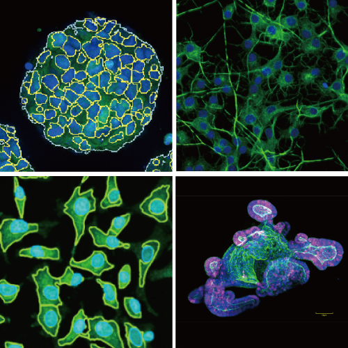
- Nipkow spinning disk confocal technology allows high-speed yet gentle 3D image acquisition
- Rich feature extraction to facilitate sophisticated cellular image analysis
- Wide field of view and tiling capability enables easy imaging of large specimen
Enables analysis of time-lapse and live-cell
- High precision stage incubator and low phototoxicity of our confocal makes the analysis of time-lapse and live-cell are possible
- Max.20fps option for fast time lapse*1
High-quality image and similar operability to a traditional flow cytometer
- Feature data and statistical graphs displayed in real-time with image acquisition
- Usable high-quality image as confocal microscope image.
- Easy to trace back to the original image from a graph spot, and make repetitive measurements
Open platform
- Connectable with external systems via handling robot*2
- Expandable to the integrated system as image acquisition and quantification instrument
- FCS/CSV/ICE data format readable by third-party data analysis software
- A variety of cell culture and sample dishes are applicable
Compact footprint, lightweight bench-top device; no need for a darkroom
| CQ1 | General fluorescent imaging | Flow cytometry | |
|---|---|---|---|
| Cell removal/suspension treatment | Not necessary | Not necessary | Necessary |
| Cell image confirmation | Possible | Possible | Not possible |
| Display feature data and graphs in real-time with imaging | Possible | Depends on devices | Possible |
| 3D data measurement | Possible | Not possible | Not possible |
| Time lapse | Possible | Not possible | Not possible |
*1 Option
*2 Contact to CQ1 partner for more information