3D CELL EXPLORER

How Nanolive’s 3D Cell Explorer Can Help You Study Living Cells
- Explore living cells without damaging them: no Stains, no fixation, no bleaching, no phototoxicity
- Obtain new insights with label-free 3D characterization of your living cells in physiological conditions without any bleaching or phototoxicity
- Perform label-free 4D continuous observation of the most sensitive cells from seconds to weeks
- Preserve the intactness of your cells: Nanolive’s 3D Cell Explorer is the world-leader in live cell assays since it delivers the highest cellular information density anywhere available on the market
- Rediscover your living cells through multiplexing: Nanolive datasets contain the simultaneous acquisition of several biological features and organelles– therefore capturing various pathways of cell biology in real-time
Rotating 3D Cell Explorer
Label-free live cell imaging of a Preadipocyte cell with Nanolive imaging
The 3D Cell Explorer’s Software
STEVE: An intuitive and comprehensive tool to explore live cell data
STEVE is the 3D Cell Explorer’s software counterpart. After the series of holograms has been captured by the hardware, high-resolution images of each plane in the sample are created by computer processing. Improved image resolution is achieved by employing a synthetic aperture and multiple-viewpoint-holographic methods.
STEVE’s intuitive interface controls the microscope, explores live cell data using interactive digital staining and even performs quantitative analysis on cell measurements. STEVE runs smoothly, even during acquisition.
Watch the video to learn more about STEVE.
Applications
Nanolive’s top applications for the 3D Cell Explorer
Neuroscience
Neuron on carpet of astrocytes.
Stem cells
High resolution imaging of mitosis in stem cells.
Immuno-Oncology
Antibodies being used as a T cell targeting system to wipe out cancer cells.
Nanolive’s holotomography
Schematic of Nanolive’s light path
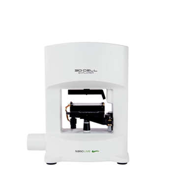
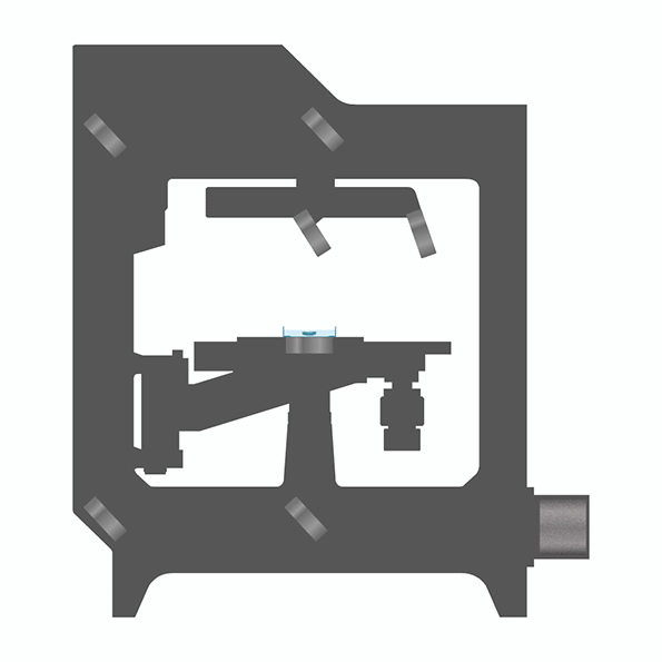
Nanolive’s unique combination of holography and rotational scanning enables high resolution imaging of cells and their organelles. This holotomographic imaging technology works label-free and reports the 3D refractive index distribution of cells and their contents. The extremely low light power that generates the holograms allows for a total absence of phototoxicity which leads to excellent time resolution.
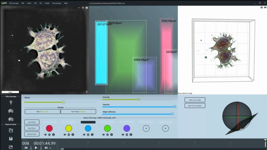
Top Image: 10h time-lapse of mESC undergoing mitosis (Refractive Index and 3D visualization). Bottom image: Organelles which are visible label-free with Nanolive technology: a. mitochondria; b. plasma membrane; c. actin fibers; d. lipid droplets; e. lysosomes; f. nuclear envelope, nucleus & nucleoli.
