CellVoyager High-Content Analysis System CQ3000

Home » Cell Imaging & Invivo Imaging » High Content Confocal Imaging » CellVoyager High-Content Analysis System CQ3000

CellVoyager High-Content Analysis System CQ3000 can acquire high-resolution 3D images at high speed while culturing cells. CQ3000 can be combined with options according to the intended use to provide a system depending on your objectives.

CQ3000 System Highlights

Excitation laser wavelength405nm、488nm、561nm、640nm
Objective lens2x、4x 、10x、20x 、40x、60x
(Dry 、Water immersion、Long working distance)
CameraHigh-sensitivity sCMOS camera (up to 2 units)
AutofocusLaser autofocus
Observation methodConfocal image, Wide-field image*1
Software (Option)High Content Analysis Software CellPathfinder

*1 Can be acquired when Uniformizer option is selected.

Introduction

In the drug discovery market, the demand for high-content analysis systems for drug efficacy evaluation screening is ever increasing year by year.

The norm has shifted from 2D to 3D and live cell evaluation.

In addition, the construction of more complex evaluation systems are sought after, including experimental systems in environments closer to the human body such as organoids that reproduce parts of various organs or organs-on-chips, as well as Cell Painting, which morphologically profiles cellular components and organelles by multicolor imaging using multiple fluorescent dyes.

Solution

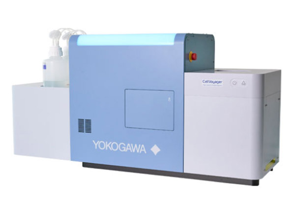
The CellVoyager High-Content Analysis System CQ3000 is a benchtop HCA system capable of acquiring high resolution images at high speed.

Yokogawa‘s proprietary technology, the Confocal Scanner Unit “CSU”, and a high-precision incubator enable stable live cell observation and high-speed imaging with low photobleaching and phototoxicity.

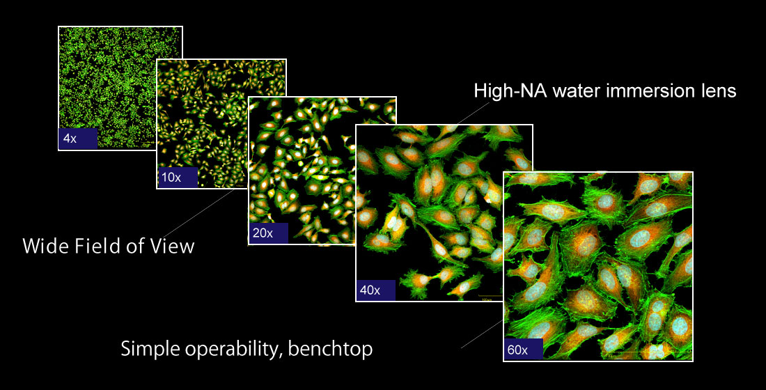
By combining options according to your application, we can provide the best system for you. Water immersion objective lens with high NA for easy observation of deep areas, uniform illumination effective for tile imaging such as tissue sections, and Target Search for automatic imaging of samples matching the conditions, all contribute greatly not only to improving the quality of experiments but also to speeding up and automating them.

In combination with the dedicated analysis software CellPathfinder, which supports Deep Learning, the system can support complex analysis and display results in graph form.

Make way for high-performance HCA optimized for laboratories!

High NA immersion lens for high quality imaging, Uniformizer for uniform and efficient imaging of the entire FOV, and a second camera for simultaneous imaging of two channels can be selected according to the application.

High throughput

  • Fast 3D imaging with YOKOGAWA’s scanning method
  • Simultaneous dual-wavelength imaging with a second camera
  • Fast 100-fps option for high-speed applications such as myocardial and calcium firing

High definition images

  • High NA water immersion lens for bright, deep imaging
  • High quantum efficiency sCMOS camera
  • The “Uniformizer”, for uniform illumination across all corners of the image

Live-cell imaging

  • Top-class incubator performance achieves stable live cell imaging
  • Incubation time of up to 7 days
  • CSU  for low bleaching and low phototoxicity imaging

Automation

  • Scans and automatically captures samples that match the conditions
  • Support for various interfaces (APIs) for external collaboration
  • Output of data types for easy integration, such as OME-TIFF and Omero